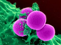
Lymph node follicles (top) and the intestinal worm Heligmosomoides polygyrus bakeri (bottom).Credit: © N. Harris (EPFL)
In order to fight invading pathogens, the immune system uses “outposts” throughout the body, called lymph nodes. These are small, centimeter-long organs that filter fluids, get rid of waste materials, and trap pathogens, e.g. bacteria or viruses. Lymph nodes are packed with immune cells, and are know to grow in size, or ‘swell’, when they detect invading pathogens. But now, EPFL scientists have unexpectedly discovered that lymph nodes also contain more immune cells when the host is infected with a more complex invader: an intestinal worm. The discovery is published in Cell Reports , and has significant implications for our understanding of how the immune system responds to infections.
The discovery was made by the lab of Nicola Harris at EPFL. Her postdoc and first author Lalit Kumar Dubey noticed that the lymph nodes of mice that had been infected with the intestinal worm Heligmosomoides polygyrus bakeri had massively grown in size. This worm is an excellent tool for studying how the worm interacts with its host, and is therefore used as a standard throughout labs working in the field.
Lymph nodes have microscopic compartments called “follicles,” where they store a specific type of immune cells, the B-cells. Stored in the follicles, B-cells pump out antibodies into the bloodstream to attack invading pathogens.
The researchers found that the mouse lymph nodes were actually producing more follicles, suggesting they were producing more B-cells in response to the worm infection. Of course, this is not a simple event. Like many biological processes, it involves an entire sequence of molecular signals that result in the formation of new cells and tissue.
Find your dream job in the space industry. Check our Space Job Board »
The EPFL scientists were able to reconstruct the molecular sequence, which is fairly complex: when the mouse is infected with the intestinal worm, a “cytokine” molecule is produced. This cytokine then stimulates B-cells in the lymph nodes to produce a molecule called a lymphotoxin. The lymphotoxin then interacts with the cells that form the foundation of the actual lymph node — the so-called “stromal cells.” The stromal cells then produce another cytokine, which stimulates the production of new follicles in the lymph node.
Until now, formation of new B-cell follicles in the lymph nodes was thought to only happen just after birth. This study provides the first detailed evidence to show that this phenomenon can take place in an adult mammal. The researchers also showed that formation of new follicles is important for fighting infection as it encourages the production of more antibodies.
Unlike bacterial or viral infections, worm infections are enormously complex. “Worms are large creatures that produce a host of their own molecules upon infection,” says Nicola Harris. “Some of these molecules stimulate the host’s immune system while some others suppress it. The field is investigating every one of these molecules, but it is slow work.”
It must be noted that the new production of B-cell follicles has only been confirmed in worm infections. “We are currently looking at this effect with bacterial infections in mice,” says Nicola Harris. “Nonetheless, we are pursuing a deeper understanding of this process to see if it is involved in producing adequate antibodies in response to vaccines.”
Source: Ecole Polytechnique Fédérale de Lausanne
Journal Reference:
- Dubey LK, Lebon L, Mosconi I, Yang C-Y, Scandella E, Ludewig B, Luther SA, Harris NL. Lymphotoxin-Dependent B Cell-FRC crosstalk promotes de novo follicle formation and antibody production following intestinal helminth infection. Cell Reports, May 2016 DOI: 10.1016/j.celrep.2016.04.023


