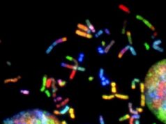
This visual abstract depicts the findings of Cortese et al., who show that Zika virus infection in both human hepatoma and neuronal progenitor cells induces drastic structural modification of the cellular architecture. Microtubules and intermediate filaments surround the viral replication factory composed of vesicles corresponding to ER membrane invagination toward the ER lumen. Importantly, alteration of microtubule flexibility impairs Zika replication. Credit: Cortese et al./Cell Reports 2017
The Zika virus taking hold of the inner organelles of human liver and neural stem cells has been captured via light and electron microscopy. In Cell Reports on February 28, researchers in Germany show how the African and Asian strains of Zika rearrange the endoplasmic reticulum and cytoskeletal architecture of host cells so that they can build factories where they make daughter viruses. The study reveals that targeting cytoskeleton dynamics could be a previously unexplored strategy to suppress Zika replication.
“Cytoskeleton elements control cellular shape, growth, and movement, provide mechanical support and stress-resilience, and coordinate organelle anchoring and vesicular transport,” says senior author Ralf Bartenschlager, a virologist at Heidelberg University. “Zika virus infections cause a drastic perturbation of the cytoskeletal network, re-organizing both intermediate filaments and microtubules into a cage-like structure that surrounds the replication machinery.”
His group, led by first author and Heidelberg postdoctoral fellow Mirko Cortese, also found that Zika and dengue, both flaviviruses, use similar strategies to infect host cells. Once inside of a cell, viral particles latch onto the rough endoplasmic reticulum—the ribosome-rich membranes outside the nucleus where RNA is translated into proteins. The viruses then remodel the endoplasmic reticulum to form a protective cage with small holes where RNA and newly made viral particles can travel in or out.
The difference with Zika is that it has unique ways of rearranging structures within the cell. For example, there are an abundance of microtubules—proteins that build the cellular cytoskeleton—surrounding its protective cage. There were also slight variations between how Zika establishes itself in human liver cells versus neural stem cells, where it is more physiologically relevant. This suggests there are yet-unknown cell-type-specific factors that affect how the virus replicates.
Find your dream job in the space industry. Check our Space Job Board »
“The cytoskeleton has a crucial role in neurogenesis,” Cortese says. “Thus, it is tempting to speculate that the alterations induced by Zika virus on the host cytoskeleton might be connected to the underlying physio-pathological mechanisms that link congenital Zika virus infections to microcephaly and neurodegenerative disorders.”
Seeing Zika’s reliance on the microtubules for its replication, the researchers now want to explore whether these drugs, including taxanes routinely used during chemotherapy, can have potential anti-viral activity against Zika in animal models. These, and similar drugs, have been declared safe to use during the second and third trimester of pregnancy, as well as during breastfeeding.
“Having identified a link between cytoskeleton dynamics and Zika virus replication, together with the plethora of available cytoskeleton-targeting compounds, might contribute to gain mechanistic insight into the ZIKV replication cycle and identify new avenues for treatment,” Bartenschlager says.
Source: Cell Press
Research Reference:
Cell Reports, Cortese et al.: “Ultrastructural characterization of Zika virus replication factories” http://www.cell.com/cell-reports/fulltext/S2211-1247(17)30179-1 , DOI: 10.1016/j.celrep.2017.02.014


