A team of University of Alberta engineers is refining a new imaging technique that could reduce the number of repeat surgeries patients undergo to remove cancerous tumors.
The team, led by Roger Zemp, is using ultraviolet photoacoustic remote sensing microscopy (UV-PARS) to rapidly visualize and analyze tumor tissue while patients are still on the surgical table, to more effectively check whether the entire tumor has been removed and ensure no repeat surgeries are needed.
The current medical practice to determine whether an entire tumor has been removed during surgery, called histology, involves pathologists examining a section of the removed tumor through a microscope in a lab, a process that can take weeks.
“They might only look at less than 0.1 percent of the volume of the tumor,” said Zemp, who is also a member of the Cancer Research Institute of Northern Alberta. “Nevertheless, histology is currently the gold standard for determining if surgeons got all the tumor out or not, to know if the margins around the lump are clean and free of tumor.”
The UV-PARS technology provides histology-like images that are comparable to the previously obtained diagnostic data, without the need to send the tissue away for analysis.
Find your dream job in the space industry. Check our Space Job Board »
Zemp’s team has developed a table-top model but is working on refining the technology to increase the speed at which it can analyze tissues and creating a smaller model that would be more suitable for potential clinical use within the next few years.
The team is also working on making the technology safe for use in vivo, which means in a living human or animal, and not just on tissue samples. Ultraviolet light, as used in UV-PARS, is not safe for general use on live tissue unless it is for a procedure in which layers of tissue are being removed, which, however, is common in surgeries.
“We have new data demonstrating in vivo imaging, and believe we really have advantages over most any other technique for virtual histology,” Zemp said.
Zemp and his team were recently awarded a grant from the Canadian Institutes of Health Research and have other pending grants to look at the technology using mid-infrared light instead, which would be safe for in vivo use. The technology has also been spun off to a startup company, illumiSonics Inc.
The first studies, “Ultraviolet Photoacoustic Remote Sensing Microscopy” and “Reflective Objective-Based Ultraviolet Photoacoustic Remote Sensing Virtual Histopathology,” were published in Optics Letters, first authored by Nathaniel Haven from the Zemp lab.
Provided by: University of Alberta
More information: Nathaniel J. M. Haven et al. Ultraviolet photoacoustic remote sensing microscopy. Optics Letters (2019). DOI: 10.1364/OL.44.003586
Nathaniel J. M. Haven et al. Reflective objective-based ultraviolet photoacoustic remote sensing virtual histopathology, Optics Letters (2019). DOI: 10.1364/OL.382415
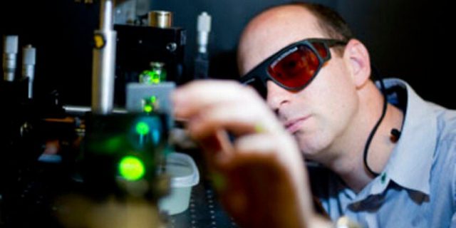
Image: U of A biomedical engineer Roger Zemp is leading research on imaging technology that uses ultraviolet light and sound to show surgeons whether they’ve completely removed cancerous tumours while the patient is still on the operating table.
Credit: Jimmy Jeong


