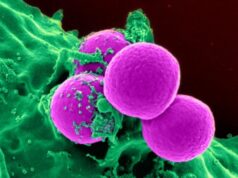
A scanning electron microscope image of Group A Streptococcus (orange) during phagocytic interaction with a human neutrophil (blue). Credit: NIAID
The human mouth can harbour more than 700 different species of bacteria. Under normal circumstances these microbes co-exist with us as part of our resident oral microbiota. But when bacteria spread to other tissues via the blood stream, the results can be catastrophic.
Researchers from the University of Bristol have now revealed a potentially key molecular process that occurs in the case of infective endocarditis, a type of cardiovascular disease in which bacteria cause unwanted blood clots to form on heart valves. If untreated, this condition is fatal and even with treatment, mortality rates remain high (up to 30 per cent). There are over 2,000 cases of infective endocarditis in the UK annually and the incidence is rising.
The Bristol team’s findings could lead to the development of new drugs to help combat this life threatening heart disease.
A key part of the study involved use of the UK national synchrotron facility, Diamond Light Source. Using this giant X-ray microscope the team were able to visualise the structure and dynamics of a protein called CshA which, based on previous studies at Bristol University, was believed to play an important role in targeting the oral bacterium Streptococcus gordonii to the tissues of the heart. The researchers were intrigued to find that CshA acts as a ‘molecular lasso’ to enable S. gordonii to bind to the surface of human cells. Such adhesive interactions are critical first steps in the ability of this bacterium to cause disease.
The study, which appears as ‘Editors’ Picks’ in the current issue Journal of Biological Chemistry, was conducted in collaboration with Professor Rich Lamont at the University of Louisville, USA.
Find your dream job in the space industry. Check our Space Job Board »
Lead author Dr Catherine Back from Bristol’s School of Oral and Dental Sciences, said: “What our work has revealed is a completely new mechanism by which S. gordonii and related bacteria are able to bind to human tissues. We have named this the ‘catch-clamp’ mechanism.”
The team were able to demonstrate that the terminal portion of CshA is very flexible. This allows it to be cast out from the surface of the bacterium like a lasso. When the lasso contacts fibronectin on the surface of human cells (the ‘catch’), it brings CshA and fibronectin into close proximity. This then enables another portion of CshA to tightly ‘clamp’ the two proteins together, anchoring S. gordonii to the host cell surface.
Co-researcher Dr Paul Race from Bristol’s School of Biochemistry and the BrisSynBio Research Centre, said: “What is particularly exciting about this work is that it opens up new possibilities for designing molecules that inhibit either the ‘catch’ or the ‘clamp’ steps in this process, or potentially both. The latter possibility is particularly intriguing, as bacteria are generally less likely to become resistant to agents that target multiple steps in an infective process.”
Dr Angela Nobbs, from the School of Oral and Dental Sciences, who co-led the study with Dr Race, added: “With the molecular level insight that our study provides, it is now a realistic possibility that we can begin to develop anti-adhesive agents that target disease-causing Streptococcus and related bacteria.”
Source: University of Bristol
Research Reference:
‘The Streptococcus gordonii adhesin CshA binds host fibronectin via a catch-clamp mechanism’ by Catherine R. Back et al in Journal of Biological Chemistry


